
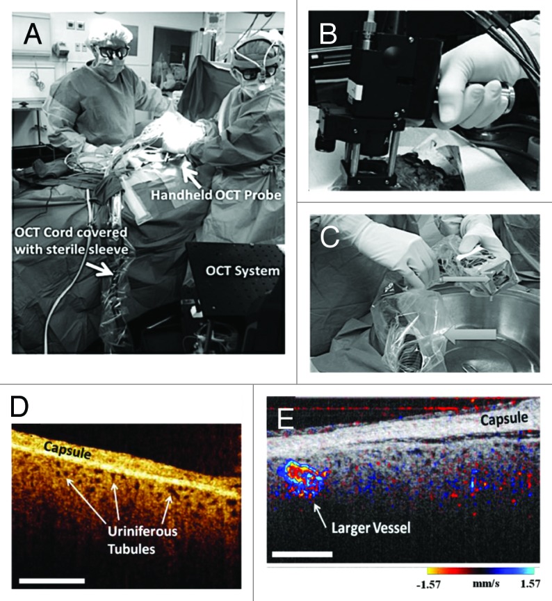
Figure 8. (A) Transplant surgeons used the sterilized hand-held OCT probe shown in (B) to image a transplanted human donor kidney in the operating room. Both surgeons are looking at real-time images of the functioning kidney. The OCT probe and associated wires are covered with a sterile camera sleeve. (C) The OCT imaging probe covered with transparent Tegaderm (small arrow). The cords leading to the probe are covered with a sterile camera sleeve (large arrow). (D) In vivo OCT imaging of human kidneys following transplantation showing open uriniferous tubules below the renal capsule. Tubules appear to be fairly open and round with some degree of homogeneity throughout the images. Scale bar is 500 μm. (E) In vivo human kidney showing open tubules and cortical blood flow. Open tubules appear round and relatively uniform across all images. Also, a larger blood vessel is seen. Scale bar is 500 μm. D and E are from reference 126 with permission.