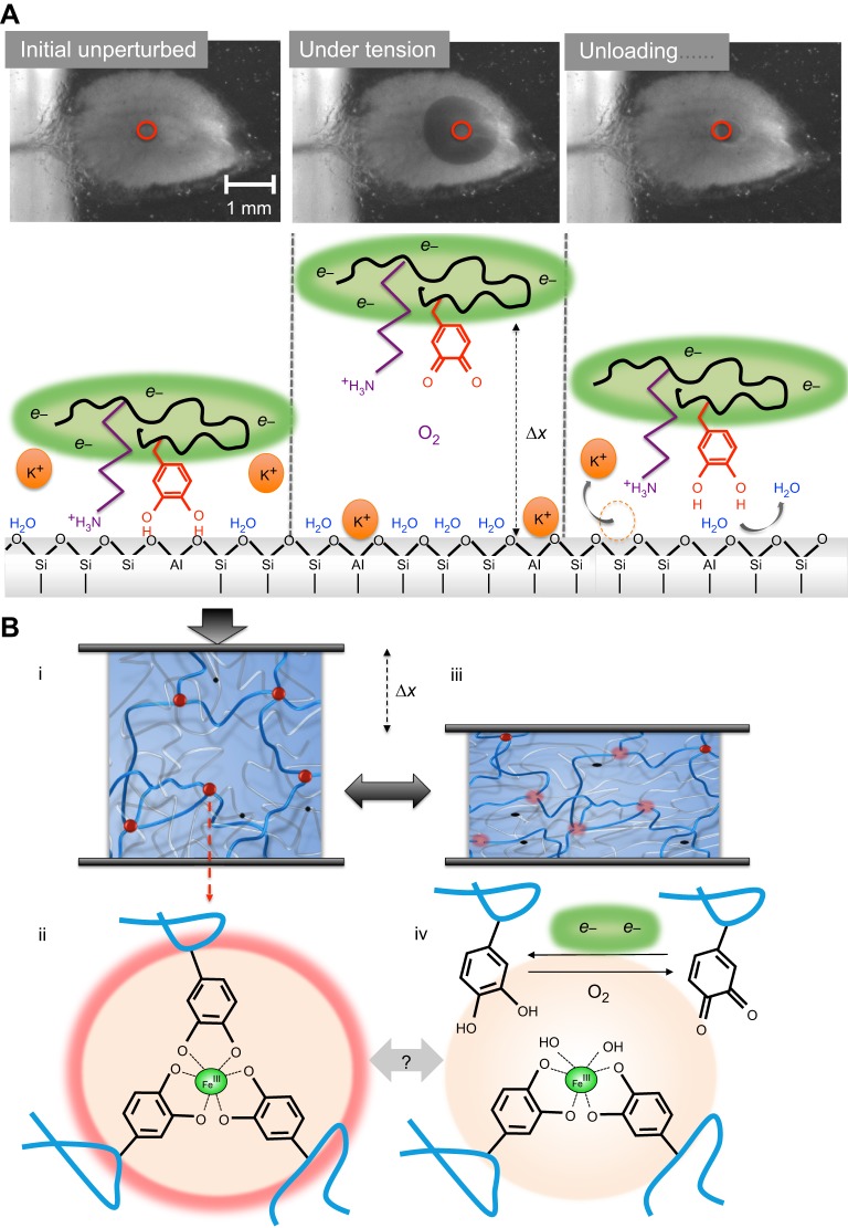
Fig. 7.
Model of chemical adaptations during cyclic loading of the plaque. (A) Interfacial events as viewed from below using transparent mica. Initial unperturbed: the interface is intact, the substratum is dry, hydrated cations (K+) are desorbed, Dopa is engaged by bidentate H- or coordination bonding, and the local environment is strongly reducing (green, e−). Under tension: upon loading, bidentate Dopa and other interactions are debonded from the surface, the interface is displaced from the substrate by some distance Δx and the surface is reinvaded by O2, salt and H2O. Oxidation of Dopa (shown) and/or thiols follows and is repaired by the reducing e− reservoir. Unloading: upon unloading, material relaxes, cations are evicted and the surface is dehydrated by Dopa, which rebonds to bidentate sites as under initial conditions. (B) Cohesive events. (i) The scheme of a double polymer network such as that in the byssal cuticle or plaque. The red balls denote tris-catecholato–iron cross-linking sites (ii) that unite three stiff (blue) Mfp-1 chains, for example. The black dots are covalent cross-linking sites that unite two compliant (silver) chains. (iii) During tensile or compressive deformation (gray arrows) by Δx (shown in compression), the tris links are at least partially disrupted (pale pink balls) allowing load transfer from stiff blue chains to compliant silver chains (iv). During disruption, catechols (Dopa) become prone to oxidation. Self-healing depends on the repair of oxidative damage. Dopa-quinone could go on to become covalent cross-links leading to embrittlement.

