Fig. 4.
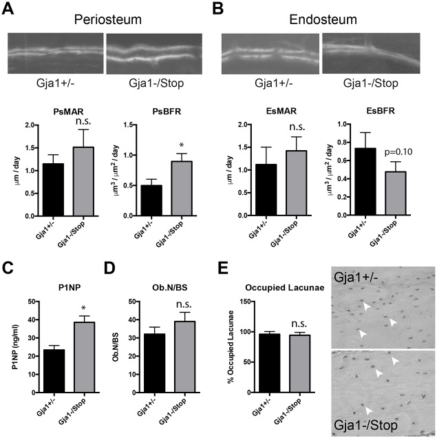
Substantial periosteal bone apposition contributes to the cortical expansion of the mid-diaphysis in 6-week-old male Gja1−/K258Stop mice. Calcein and Alizarin dual labeling and quantification of the mineral apposition rate (MAR) and bone formation rate (BFR) is shown for each genotype at the (A) periosteal (Ps) and (B) endosteal (Es) surface of the femoral diaphysis (n=3 animals per genotype, 3 sections per animal). (C) Quantification of serum levels of P1NP (n=4 per genotype). (D) Static histomorphometry of osteoblast number normalized to the bone surface area (n=5 per genotype). (E) Quantification of the percentage of occupied osteocyte lacunae observed in cortical bone from the indicated genotype (n=3 animals and >1000 lacunae/genotype). A representative hematoxylin-stained image of cortical bone is shown. White arrowheads point to occupied lacunae. Graphs depict mean±s.d. *P<0.05; n.s., not significant (two-tailed t-test).

