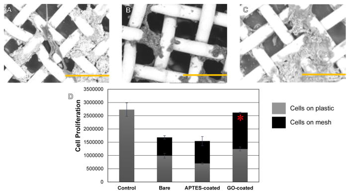
Figure 6.
Representative SEM images of SH5YSY cells grown atop GO-coated meshes in (A) (B) and (C). Depicted scale bars are a 100 μm. (D) Confirmation of cell proliferation on GO-coated meshes. Significantly greater no. of viable proliferating cells were detected on GO-coated meshes after 72 hr of culture compared with bare and APTES-coated meshes.

