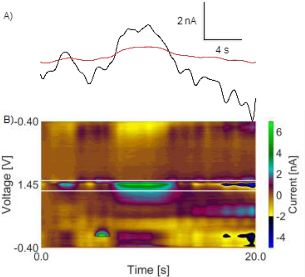
Figure 5.
in vivo stimulated histamine from the premammillary nucleus. A) Primary (black) and secondary (red) oxidation peak i vs t traces of stimulated histamine fail lag time filter because maximums occur at the same time. B) False color plot of stimulated histamine. White lines are pmax and smax generated from adenosine transients.

