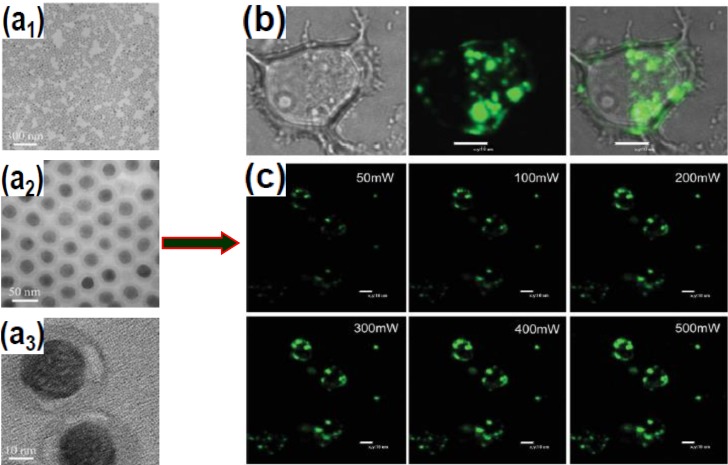
Figure 8.
Silica-coated NaYF4:Yb/Er nanocrystals and their application for cell imaging. (a1–Ca3) TEM images of silica-coated NaYF4:Yb/Er UCNPs upconversion nanoparticles at different magnifications; (b) Confocal fluorescence image of MCF-7 cells using silica-coated NaYF4:Yb/Er nanospheres (Left: bright-field, middle: upconversion image under 980 nm excitation, and Right: superimposed images of MCF-7 cells incubated with the nanoparticles for 24 h); (c) Confocal fluorescence images of MCF-7 cells with the nanospheres, excited by a 980 nm laser with different power intensities. Reproduced from [103]. Copyright 2008, John Wiley and Sons.

