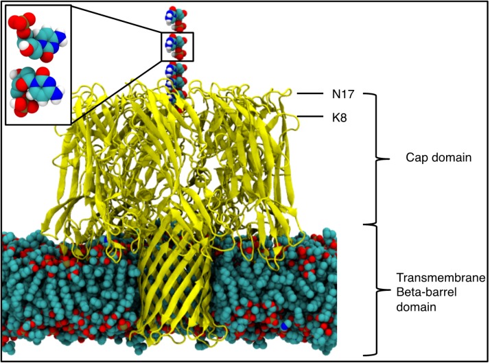
Figure 1.
The alpha-hemolysin (αHL) protein in a 1,2-dimyristoyl-sn-glycero-3-phosphocholine (DMPC) bilayer, with the mononucleotide positioned above the vestibule entrance, where each mononucleotide represents an individual simulation. The protein is represented by yellow ribbons and for other atoms: Carbon is shown in cyan, oxygen in red, nitrogen in blue, phosphorus in brown and hydrogen in white. The waters and ions are excluded for clarity. (Inset) The phosphate orientations used, termed the “up” and “down” orientations, shown top and bottom respectively.

