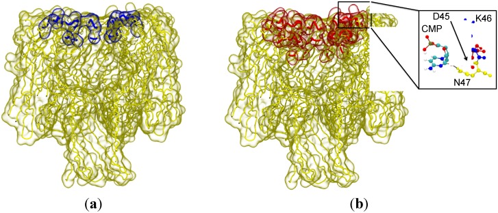
Figure 3.
The residues with the highest propensity to interact with the nucleotide for: failed capture (a) and possible capture (b). The blue highlighted residues corresponding to failed capture are on the edge of the vestibule entrance and on the surface of the vestibule. The red highlighted residues associated with possible capture are predominantly on the edge of the entrance to the vestibule. (Inset) The most frequently observed binding mode between cytosine monophosphate (CMP) and the amino acid triplet, D45, K46 and N47. Hydrogen bonds (dashed lines) are observed between the hydroxyl group of the sugar and the side-chain of D45, as well as the nucleobase and the side-chain of N47. These hydrogen bonds are stable and are present for extended periods of time (greater than 1 ns). Residues are coloured for clarity; D45 in red K46 in blue, and N47 in yellow.

