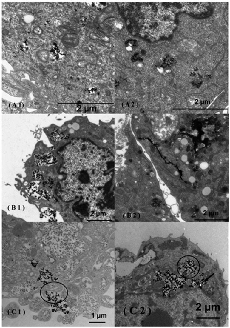
Figure 1.
Transmission electron microscopy (TEM) images of MCF-7 (A1,A2), 3T3 (B1,B2) and Caco-2 (C1,C2) cells incubated in the presence of uncoated super-paramagnetic iron oxide NPs (SPIONs). The uncoated SPIONs were internalized via endocytosis in the Caco-2 cells (C1) and afterward released into the cytoplasm (C2). Citrate-coated SPIONs agglomerated in the cytoplasm of MCF-7 cells (A2) and were adsorbed along the endoplasmic reticulum in 3T3 cells (B2). Reproduced with permission from [17]. Copyright 2014, ACS Publications.

