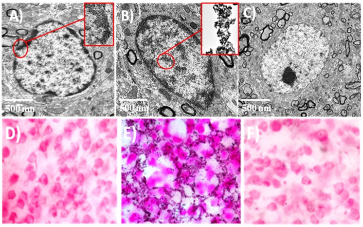
Figure 5.
TEM micrographs for the apoptotic cells before (A) and after (B) injection of SR-FLIVO-FNPs. (C) control cell in non-ischemic lesion. Light microscope images after Prussian blue staining for (D) control rat brain after injection of SR-FLIVO-FNPs, (E) ischemic lesion of rat brain with localized SR-FLIVO-FNPs, and (F) non-ischemic lesion of rat brain after injection of SR-FLIVO-FNPs.

