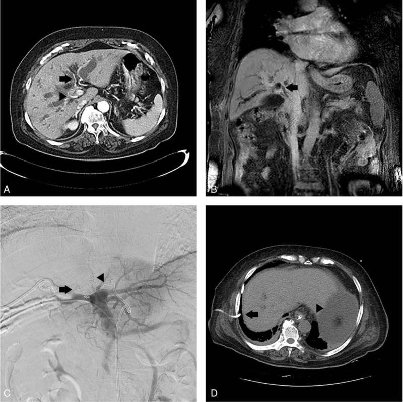
Figure 1.

(A) Case 1 patient's axial view of abdominal CT, performed prior to the PVE procedure, reveals bilateral IHD dilatation, enhanced wall thickening of hepatic duct, abutting S4 hepatic artery (arrow), and encasing right hepatic artery (arrow head). Segments II and III FLR was 29%. (B) Coronal view of preprocedure abdominal MRI reveals enhanced wall thickening at hepatic duct, CHD, and CBD to just intrapancreatic portion (arrow). (C) Trisectional PVE was performed using gelfoam particles and interlock coils with a diameter of 5 mm. Right PVE was performed (arrow) after the segment IV portal vein (P4) was blocked (arrowhead). (D) Noncontrast abdominal CT, performed 8 days post-PVE, shows the right-sided PTBD (arrow) dislocated into the abdomen and ascites (arrowhead). The segments II and III, that is, FLR, was 29.5%, with a 0.5% increase in volume. CBD = common bile duct, CHD = common hepatic duct, CT = computed tomography, FLR = future liver remnant, IHD = intrahepatic duct, MRI = magnetic resonance imaging, PTBD = percutaneous transhepatic biliary drainage, PVE = portal vein embolization.
