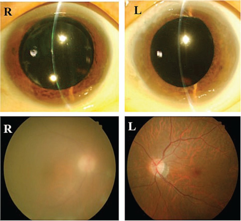
Figure 2.

Eyes during recurrence of HTLV-1 uveitis. Top left: mutton-fat keratic precipitates are present in the anterior of the right eye. Bottom left: vitreous opacity is present, and fundus visibility is poor. Right column: no inflammation is evident in the left eye. HTLV-1 = human T-lymphotropic virus type 1.
