
Figure 2.
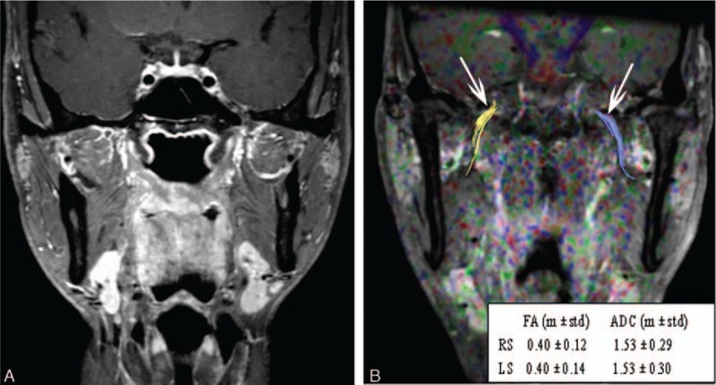
A routine MR image and fused DTI and turbo field echo (TFE) sequence images of a 42-year-old man who was a healthy control. (A) Routine coronary magnetic resonance images with fat suppression and contrast enhancement show no lesion in the nasopharynx or paranasopharyngeal space. (B) A fused DTI and TFE sequence image. Diffusion tensor tractography of the V3 branch of the trigeminal nerve was generated bilaterally (white arrows). The FA and ADC values showed no significant difference between the right side (RS) and the left side (LS).