Figure 4.

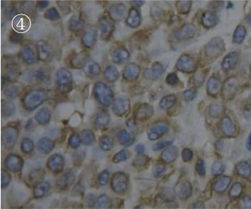
The photomicrograph of vimentin immunohistochemical staining. It was shown that the tumor cells were positively stained with vimentin antibody (magnification, ×400).

The photomicrograph of vimentin immunohistochemical staining. It was shown that the tumor cells were positively stained with vimentin antibody (magnification, ×400).