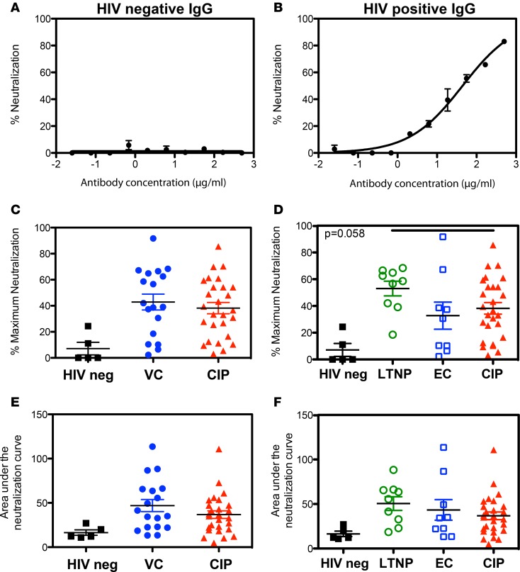
Figure 5. IgG from viremic controllers (VCs) and chronically infected patients (CIPs) do not differ in their HIV-1 neutralizing activity.
(A and B) Controls for HIV-1 neutralization assay carried out using TZM-bl cells infected with X4-tropic HIV-1 (pNL4-3). Neutralization curve for (A) polyclonal HIV– IgG sample or (B) polyclonal HIV+ IgG sample are shown. For HIV-1 neutralization assays, polyclonal IgG were diluted 3-fold starting from a top final concentration of 500 μg/ml in a 10-point titration curve. (C) Maximum neutralization activity of patient-derived IgG comparing noninfected (HIV neg) with VC and CIP. (D) Maximum neutralization activity with VCs split into LTNP and EC subgroups. (E) Neutralization activity as measured by the area under the neutralization curve (AUC) for IgG purified from CIP, VC, and noninfected donors (HIV neg). (F) Neutralization activity (AUC) with VCs split into LTNP and EC subgroups. The mean of 3 experiments for n = 50 IgG is shown.

