
FIGURE 1.
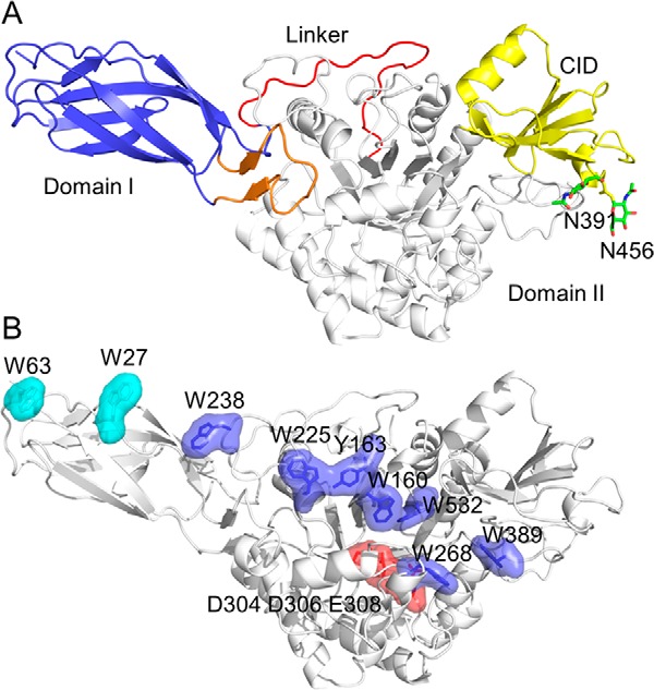
Structure of OfChi-h. A, schematic representation of OfChi-h. Domain I is shown in light blue, domain II is shown in white, chitinase insertion domain (CID) from domain II is shown in yellow, the linker is shown in red, and the motif that contributes to the domain I-domain II interaction is shown in orange. N-Glycan sites are shown as green sticks. B, surface representation of OfChi-h. The solvent-exposed aromatic residues in domain I (Trp27 and Trp63) and domain II (Trp160, Tyr163, Trp225, Trp238, Trp268, Trp389, and Trp532) are shown in cyan and blue, respectively. The catalytic residues (Asp304, Asp306, and Glu308) are shown in red.