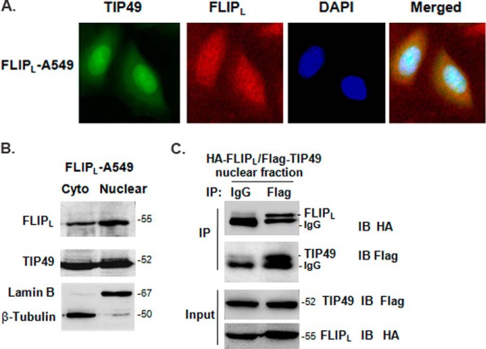
FIGURE 2.
The nuclear localization of c-FLIPL and TIP49. A, endogenous TIP49 and c-FLIPL were detected by immunofluorescence in FLIPL-A549 cells, respectively. Merged presents the images overlapped. B, the cytoplasmic (cyto) and nuclear fractions of FLIPL-A549 cells were prepared using the Nuclear and Cytoplasmic Protein Extraction Kit and detected by antibodies as indicated. Lamin B and Tubulin were used as nuclear- or cytoplasmic-specific protein loading controls, respectively. C, 293T cells were cotransfected with HA-FLIPL and FLAG-TIP49 expression vector. After 24 h transfection, cells were prepared for nuclear extraction. Nuclear pellets were resuspended in lysis buffer and immunoprecipitated with IgG or anti-FLAG antibody and probed with anti-HA antibody.

