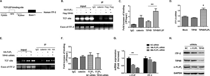
FIGURE 5.
The role of c-FLIPL-TIP49 involved in the β-catenin·TCF transcriptional complex. A, schematic representation of the TCF binding site in the human ITF-2 promoter at −1874. Forward (F) and reverse (R) PCR primers were used to detect the ITF-2 promoter region in the ChIP assay. B, 293T cells were transfected with FLAG-TIP49 or cotransfected with HA-FLIPL/FLAG-TIP49. After 24 h transfection, cells were harvested for ChIP assay using antibodies as indicated. ChIP DNA was analyzed with a pair of primers that flank the TCF binding site at −1874. As negative controls, primers corresponding to a downstream coding region of ITF-2 were used. Input DNA used for PCR corresponded to 0.1% of total chromatin DNA for immunoprecipitation. Reactions were assessed by regular PCR (B) and qPCR (C). D, ITF-2 mRNA expression was detected by qPCR in transfected cells. GAPDH was used as housekeeping gene controls. E, 293T cells were first transfected with siRNAs for negative control or TIP49 for 24 h, and then transfected with HA-FLIPL expression vector for an additional 24 h. ChIP assay was performed using antibodies as indicated. Reactions were assessed by regular PCR (E) and qPCR (F). G, ITF-2 mRNA was detected by qPCR when cells were treated with siRNAs for c-FLIPL. H, the protein level of ITF-2 was detected by Western blot analysis when knocking down c-FLIP or TIP49.

