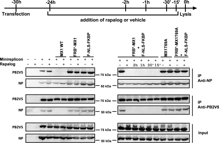
FIGURE 7.
Activated FRB*-MX1 disrupts the interaction between PB2V5 and NP. HEK293T cells were transfected with PB1, PB2V5, PA and NP expression plasmids and pHW-NSLuc (100 ng each). The cells were co-transfected with 1 μg of expression vector for MX1 WT, MX1T69A, FRB*-MX1, and F-NLS-FKBP and 0.5 μg of NP expression vector as indicated. The cells were treated with 100 nm rapalog at different time points after transfection as indicated. Vehicle was added as a negative control for the −24 h time point (−). Cells were lysed 30 h after transfection. PB2V5 and NP were immunoprecipitated (IP) with anti-V5 and anti-NP antibodies, respectively. Proteins in the NP (IP anti-NP) and PB2V5 (IP anti-V5) immunoprecipitates and total cell lysates (input) were visualized by Western blotting with antibodies specific for NP (anti-RNP antibody) and PB2V5 (anti-V5 antibody). The result shown is representative of three independent experiments.

