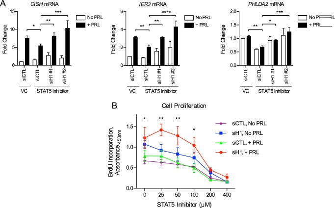
FIGURE 9.
H1 knockdown enhances gene expression and breast cancer cell proliferation in response to reduced STAT5 activation. A, H1 knockdown rescues gene expression following partial STAT5 inhibition. T47D cells were transfected with siCTL, siH1-1, or siH1-2 (Fig. 8A). Transfectants were pretreated with the STAT5 inhibitor (200 μm) or DMSO VC for 1 h, followed by PRL treatment for 2 h. RNA was isolated, and cDNA was synthesized by RT-PCR and analyzed by qPCR. -Fold change was calculated relative to VC, siCTL with no PRL treatment. Results are presented as the mean ± S.E. (error bars), n ≥ 3 independent experiments. Statistical significance was determined by a two-sided ratio paired t test. B, H1 knockdown rescues cell proliferation in response to intermediate levels of STAT5 inhibition. T47D cells were transfected with siCTL or siH1 (pooled 1 and 2). Transfectants were treated with the indicated concentrations of STAT5 inhibitor, with or without PRL, for 3 days. BrdU incorporation was measured by absorbance as an indication of cell proliferation. Results are presented as the mean ± S.E. of three independent experiments. Within each individual experiment, each set of treatment conditions was carried out in triplicate. Statistical significance was determined by two-sided t test assuming equal sample variance. Statistical significance shown in the figure is comparing siCTL + PRL versus siH1 + PRL at the indicated concentration of STAT5 inhibitor. Other statistically significant comparisons are as follows: siCTL No PRL versus siH1 No PRL (p ≤ 0.01 at 0 inhibitor; p ≤ 0.05 at 25 μm); siH1 No PRL versus siH1 + PRL (p ≤ 0.05 at 25 μm; p = 0.052 at 50 μm). *, p ≤ 0.05; **, p ≤ 0.01; ***, p ≤ 0.001; ****, p ≤ 0.0001.

