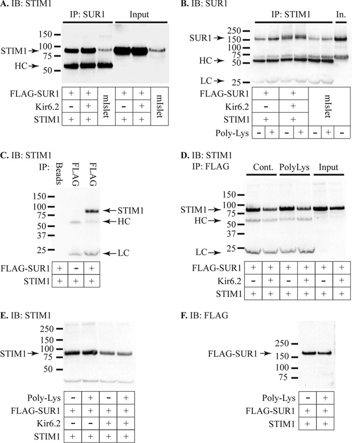
FIGURE 7.
Interaction of STIM1 and KATP. A, IP studies using mouse islet (mIslet) extracts or HEK293T cell extracts expressing STIM1 and FLAG-SUR1 in the presence or absence of Kir6.2. IP was performed using anti-SUR1 antibody followed by IB with anti-STIM1.The SUR1 antibody pulled down STIM1 in all three samples. The input protein (IB only with anti-STIM1) is shown in the right three lanes. The band indicated as HC represents the heavy chain of the IP antibody, detected by the secondary antibody, and is only seen in the three IP samples. Co-immunoprecipitation was observed in two independent experiments using mouse islet or HEK cell proteins performing IP with anti-STIM1 and IB with anti-SUR1. B, IP was performed using anti-STIM1 and IB with anti-SUR1 using the same protein samples as in A, representative of two independent experiments with this protocol. The STIM1 antibody pulled down SUR1 in all three samples, and pulldown was enhanced by adding 50 μg/ml poly-lysine to the samples during IP. The SUR1 antibody exhibited a secondary band of slightly higher molecular weight than the heavy chain band, and subsequent experiments were performed using anti-FLAG to avoid any confounding effects of this band. LC represents the light chain of the IP antibody, and In is the mouse islet input protein. C, control IP studies using HEK293T cells expressing STIM1 in the presence or absence of FLAG-SUR1. IP performed with protein A-agarose beads (Beads) alone with samples expressing STIM1 and FLAG-SUR1 failed to pull down STIM1. IP with the FLAG antibody in cells expressing STIM1 but not FLAG-SUR1 also failed to pull down STIM1. IP using the FLAG antibody did, however, pull down STIM1 in cells expressing both STIM1 and FLAG-SUR1. STIM1 was detected by IB with anti-STIM1 along with nonspecific bands for the heavy-chain (HC) and light-chain (LC) of the FLAG antibody in the two FLAG-IP lanes but not the beads-only lane. IP of STIM1 using the FLAG antibody was observed in six experiments using four different protein samples. D, IP with the FLAG antibody pulled down STIM1 in the presence or absence of Kir6.2 under control conditions, and this pulldown was enhanced by the addition of 50 μg/ml poly-lysine (PolyLys). The image shown is representative of three independent experiments, and the increase in band density with poly-lysine was 27 ± 3% (n = 9, range 17 −44%) relative to control for cells expressing STIM1 and FLAG-SUR1 and 11 ± 1% (n = 6, range 8–16%) for cells expressing STIM1, FLAG-SUR1, and Kir6.2. Densities of STIM1 IB bands were significantly higher for poly-lysine-treated extracts relative to controls for these samples with STIM1 and FLAG-SUR1 (paired t test, p = 0.003) and also for samples with STIM1, FLAG-SUR1, and Kir6.2 (paired t test, p < 0.001). E, the STIM1 band density was 97 ± 2% (n = 6, range 92–102%, not significantly different, paired t test) relative to control for cells expressing STIM1 and FLAG-SUR1 and 94 ± 5% (n = 6, range 83–105%, not significantly different, paired t test) for cells expressing STIM1, FLAG-SUR1, and Kir6.2. F, the FLAG band density was significantly lower with poly-lysine (91 ± 6%, n = 10, range 75–133%, p = 0.04, three independent experiments, paired t test) relative to control for cells expressing STIM1 and FLAG-SUR1 and 83 ± 5% (n = 7, range 74–99%, p = 0.03, two independent experiments, paired t test) for cells expressing STIM1, FLAG-SUR1, and Kir6.2.

