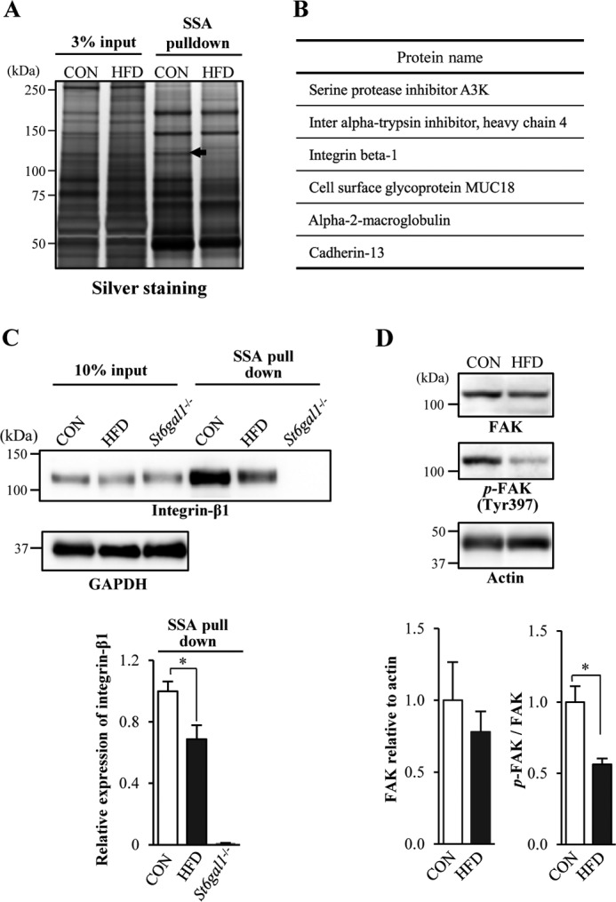
FIGURE 2.

Identification of α2,6-sialylated proteins reduced in VATs from obese mice. A, α2,6-sialylated proteins were pulled down with SSA lectin from mouse VATs and then subjected to SDS-PAGE and silver staining. A precipitated protein (arrow, 120–130 kDa) from CON mice that was reduced in HFD-induced obese mice was further analyzed. B, proteins (excised from the 120–130-kDa band) identified by MS. C, proteins extracted from mouse VATs (input) and then pulled down with SSA lectin (SSA pulldown) were blotted with anti-integrin-β1 antibody or anti-GAPDH antibody (n = 3). D, proteins in VATs from CON or HFD mice were blotted with an anti-FAK, anti-phospho-FAK (p-FAK), or anti-actin antibody (n = 3). The signal intensity of the bands in the Western blot was quantified and is shown as a graph. All graphs show mean ± S.E. (*, p < 0.05 by Student's t test).
