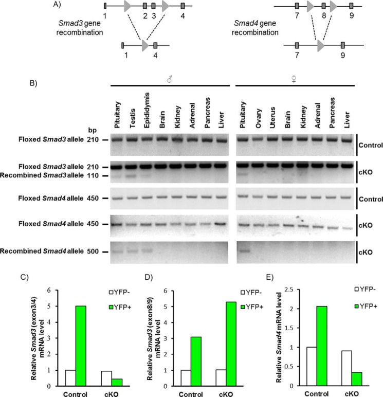
FIGURE 3.
Recombination of Smad3 and Smad4 alleles in mouse gonadotropes. A, schematic representation of the floxed Smad3 and Smad4 alleles pre- and postrecombination with Cre. Exons 2 and 3 of Smad3 and exon 8 of Smad4 were flanked with loxP sites (light gray triangles). Exons are shown as dark gray boxes. B, PCR detection of floxed and recombined Smad3 and Smad4 alleles from the indicated tissues of control and cKO mice. C–E, RT-qPCR analysis of mRNA levels of Smad3 and Smad4 in purified gonadotropes (YFP+) and non-gonadotropes (YFP−) from adult control (YFP/+;GRIC) and cKO (Smad3fx/fx;Smad4fx/fx;YFP/+;GRIC) mice. In C and E, primers were directed against the deleted region of the genes/transcripts. In D, primers were directed against non-targeted exons in Smad3.

