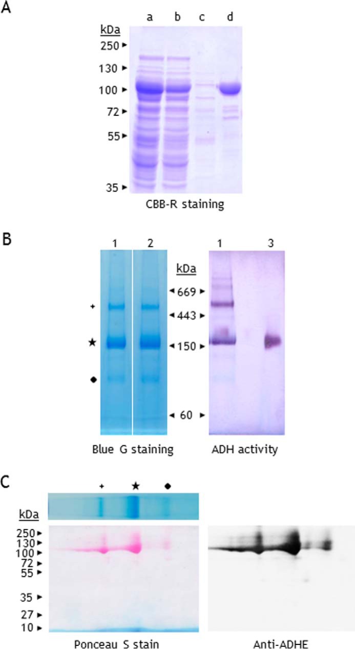
FIGURE 5.

PAGE analysis of C. reinhardtii recombinant ADHE. A, purification of His-tagged C. reinhardtii ADHE (rADHE). Coomassie Brilliant Blue R stain of proteins separated on SDS-PAGE (10%). Lane a, E. coli soluble fraction; lane b, flow-through; lane c, 10 mm imidazole wash; lane d, rADHE eluted with 100 mm imidazole (∼10 μg). B, freshly purified rADHE (25 μg) was subjected to BN-PAGE (3–12%). Lane 1, rADHE purified under anaerobic conditions; lane 2, rADHE purified under atmospheric conditions; lane 3, S. cerevisiae ADH1 (0.5 μg). C, detection of rADHE by immunoblotting after two-dimensional BN/SDS-PAGE. One BN gel lane was excised, subjected to denaturation in 1% SDS and 1% β-mercaptoethanol, and further loaded on SDS-PAGE (10%). Proteins were then transferred to a nitrocellulose membrane and stained with Ponceau S. The membrane was probed for ADHE.
