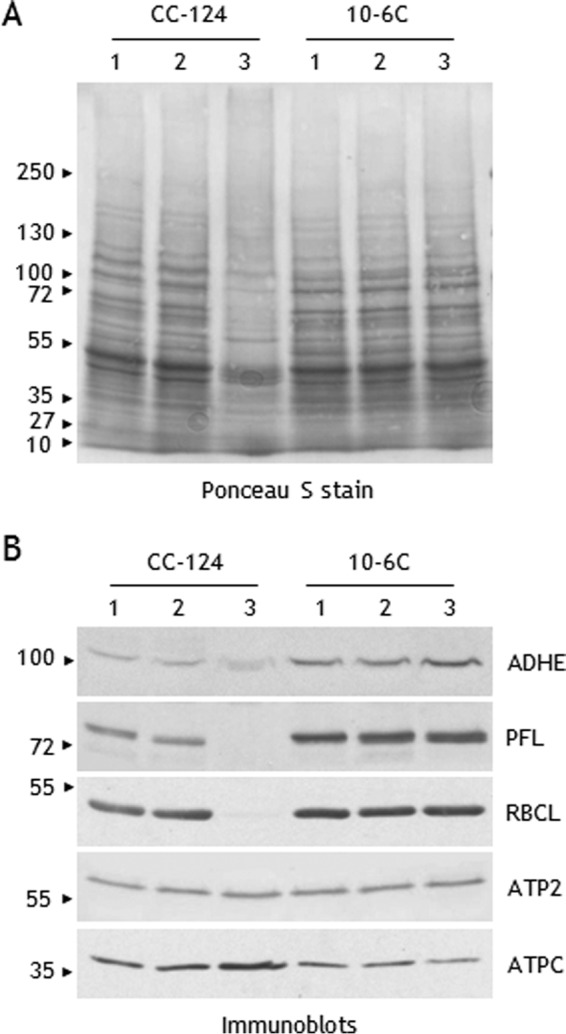
FIGURE 7.

Protein abundance in response to dark anoxia in strains CC-124 (wild type) and 10-6C (lacks Rubisco carboxylase activity). Cells were kept under dark anoxia in AIB medium at a cell concentration of 107 cells ml−1. Proteins in cell extracts (40 μg) were separated by urea/SDS-PAGE (6 m urea, 5–12% acrylamide gel) and transferred to nitrocellulose membrane. Lane 1, exponentially grown cells; lane 2, 6 h of incubation; lane 3, 24 h of incubation. A, nitrocellulose membrane was stained with Ponceau Red S. B, select proteins were detected by immunoblot analyses with antisera against ADHE, PFL, the large subunit of Rubisco (RBCL), subunit β of mitochondrial ATPase (ATP2), and subunit γ of chloroplast ATPase (ATPC). Note that strain 10-6C exhibits higher levels of ADHE and PFL.
