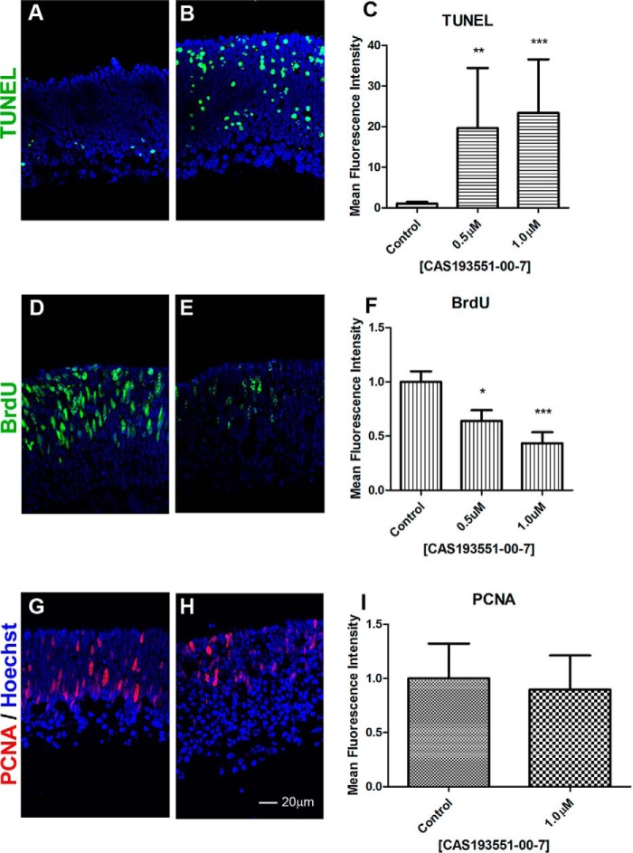
FIGURE 7.
HDAC1 inhibition reduces BrdU incorporation and increases apoptosis. Immunofluorescence microscopy of PN1 retina explants cultured for 48 h (A–F) or 96 h (G–I) with DMSO (control; A, D, and G) or treated with 1.0 μm CAS 193551-00-7 (HDAC1i; B, E, and H) was performed. Cryosections were stained with TUNEL labeling (A and B), (green), anti-BrdU antibodies for BrdU incorporation (green) (D and E), or anti-PCNA antibodies (red) (G and H), and nuclei were counterstained with Hoechst 33358 (blue). Image quantification of immunofluorescence intensity was performed for three biological replicates with three technical replicates for each sample and normalized to control ±S.E. C, TUNEL labeling. F, BrdU incorporation. I, PCNA level. *, p < 0.05; **, p < 0.01; ***, p < 0.001. Error bars represent S.E.

