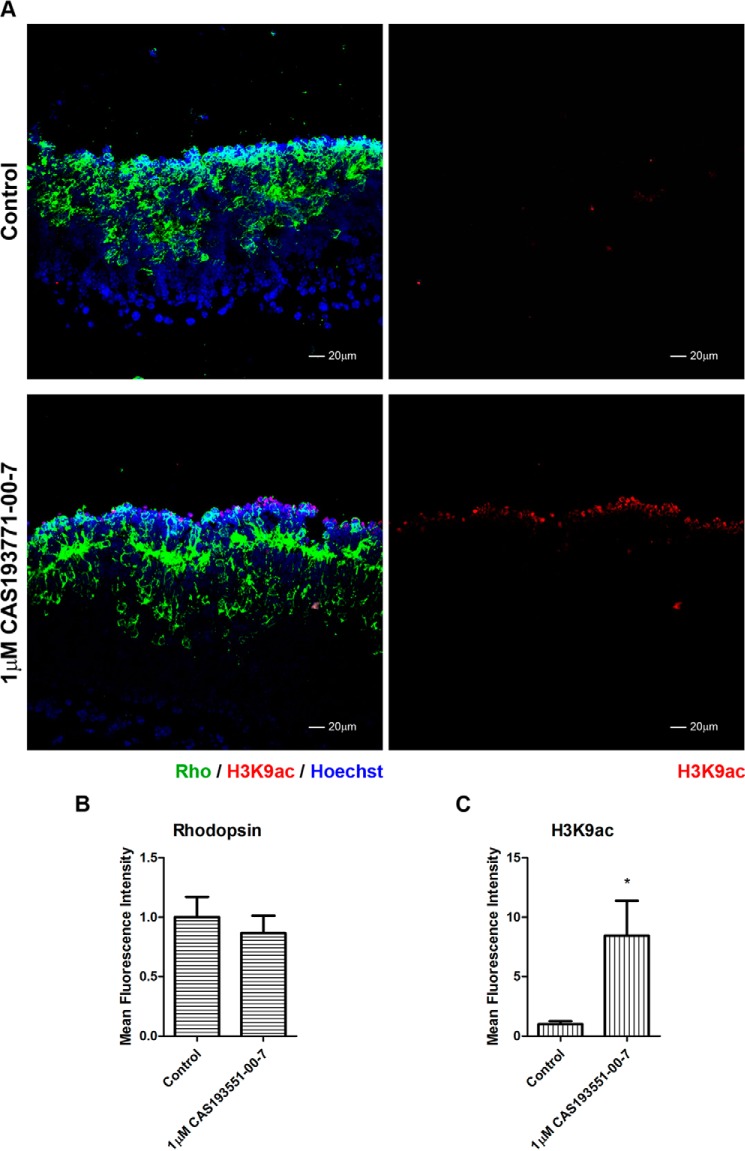
FIGURE 9.
HDAC1 inhibition of rhodopsin is stage-specific. A, immunofluorescence microscopy of PN7 retina explants cultured for 48 h with DMSO (control; upper panels) or 1.0 μm CAS 193551-00-7 (HDAC1i; lower panels). Cryosections were stained with anti-RHO (green) (left panels) and anti-H3K9ac (red) antibodies, and nuclei were counterstained with Hoechst 33358 (blue). Image quantification of immunofluorescence intensity was performed for three biological replicates with three technical replicates for each sample ±S.E. B, rhodopsin expression. C, H3K9ac levels. *, p < 0.05. Error bars represent S.E.

