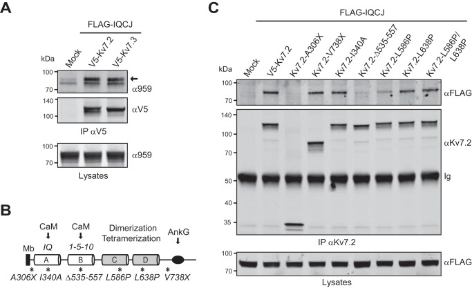
FIGURE 2.
Association of IQCJ-SCHIP1 and Kv7.2/Kv7.3 in transfected COS-7 cells. A, IP on lysates from transfected COS-7 cells expressing FLAG-IQCJ and V5-Kv7.2 or V5-Kv7.3, with V5 antibodies, and revealed by immunoblotting with V5 and SCHIP1 (α959) antibodies. Crude protein extracts (Lysates) were immunoblotted to verify protein expression. V5 antibodies co-immunoprecipitate FLAG-IQCJ. The arrow indicates the position FLAG-IQCJ detected with the α959 antibody; the lower band is not specific. B, schematic structural organization of the C-terminal intracellular domain of Kv7.2 and position of the mutations or deletions (*). Arrows indicate the regions of interaction with AnkG and calmodulin (CaM). A and B, amphipathic α-helices containing the IQ and the two adjacent 1-5-10 consensus CaM-binding motifs, respectively; C and D indicate coiled-coils; Mb, plasma membrane. C, IP on lysates from cells expressing FLAG-IQCJ and WT or mutant Kv7.2 proteins, with Kv7.2 antibodies and revealed with FLAG and Kv7.2 antibodies. FLAG-IQCJ does not co-immunoprecipitate with Kv7.2 mutants deleted of the C-terminal intracellular domain (Kv7.2-A306X) or of the CaM binding 1-5-10 motif (Kv7.2-Δ535–557). Mock, transfection with an empty vector. Molecular mass markers are shown in kDa on the left of the panels.

