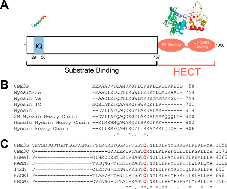
FIGURE 1.
Alignment of UBE3B with select IQ motif proteins and HECT E3 ubiquitin ligases. A, schematic of UBE3B showing the IQ domain (amino acids 29–58) and the HECT domain (amino acids 757–1068). The proposed 3D structures of the IQ and HECT domains using Phyre2 are shown above the schematic. The N terminus of HECT domains are known to bind to substrate. The HECT domain is composed of two lobes as follows: the N-lobe binds the E2(s), and the C-lobe contains the catalytic cysteine that binds ubiquitin. B, alignment of UBE3B with calmodulin binding domains as predicted by Phyre2 and using ClustalW2. C, alignment of UBE3B with HECT E3 ligase domains as predicted by Phyre2 and using ClustalW2. The conserved catalytic cysteine is highlighted in red. * denotes a single fully conserved residue; : denotes conservation between groups of strongly similar properties, . denotes conservation between groups of weakly similar properties.

