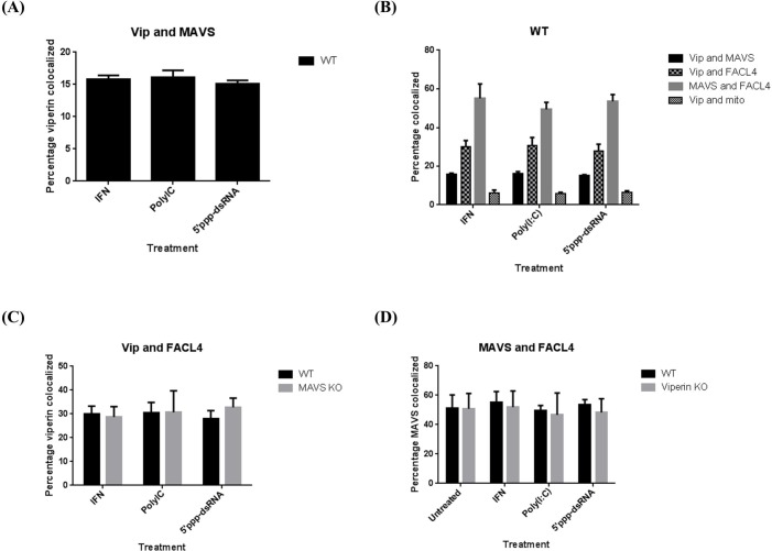
Fig 2. Viperin and MAVS do not influence each other’s intracellular distribution.
(A) WT mouse BMM were stimulated with universal type I IFN or transfected with poly(I:C) or 5’ppp-dsRNA and co-localization of viperin with MAVS quantified after 8 hrs as in Fig 1. (B) WT mouse BMM were stimulated with universal type I IFN or transfected with poly(I:C) or 5’ppp-dsRNA for 8 hours and co-localization of various marker pairs quantified. (C) WT and MAVS KO mouse BMM were stimulated for 8 hours and co-localization of viperin with the MAM marker FACL4 was quantified. (D) WT and viperin KO mouse BMM were stimulated for 8 hours and co-localization of MAVS with FACL4 was quantified. All results are presented as ±SEM of least 10 different cells.

