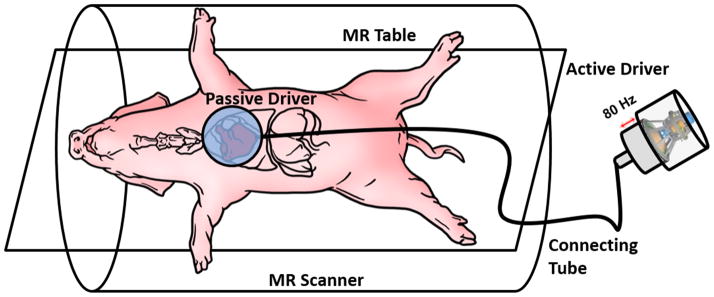
Figure 1. Experimental Set-Up.
The animal is placed feet-first supine on the MR table. A custom built passive driver is positioned externally on the animal’s anterior chest wall right above the heart. A custom made active pneumatic driver that is placed outside the scanner room generates acoustic waves and transmits it to the passive driver via a plastic connecting tube.

