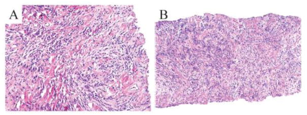
Figure 4. Pathologic analysis of needle core biopsy is consistent with mammary myofibroblastoma.
The tumor comprises of neoplastic spindle cells that are interspersed by bands of collagen. The neoplastic cells display bland nuclei and eosinophilic cytoplasm. No mitotic figures are evident (A and B; H&E).

