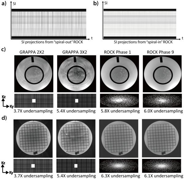
Figure 3.
The SI projections from the “spiral-out” ROCK (a) is subject to distortions due to eddy current artifacts and the same artifact is much alleviated in the “spiral-in” ROCK (b). Compared with images acquired using GRAPPA under-sampling, the images acquired using ROCK in 1.5T (c) and 3T (d) have better quality and less artifacts, though high under-sampling rates were achieved. In the ROCK method, the k-space after retrospective data binning has near-uniform angular distributions. The slightly different sampling pattern and under-sampling rate of different cardiac phases do not result in noticeable differences in image quality.

