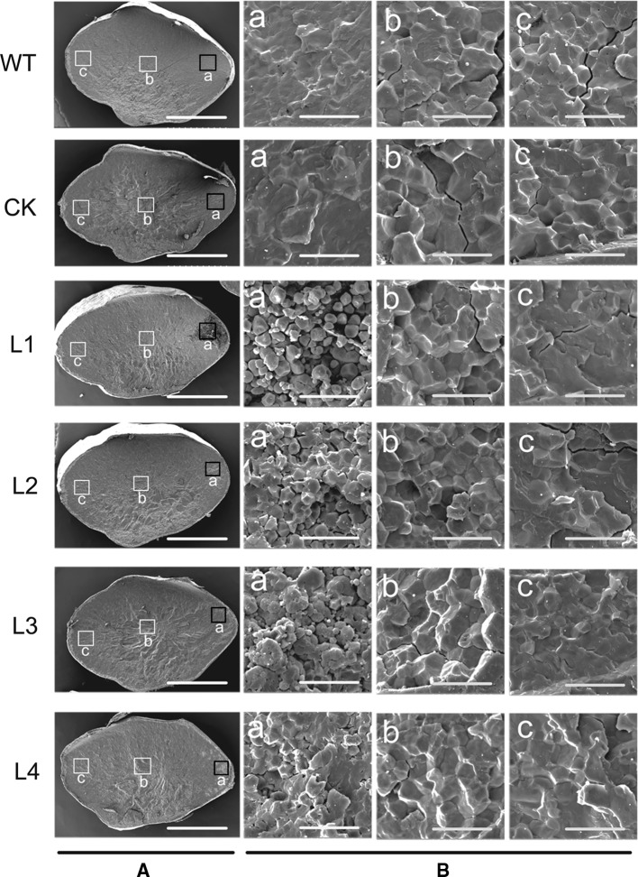
Fig. 8.
Scanning electron microscopy (SEM) images of the transverse sections of rice seeds. Cross-sections of mature seeds are shown in A. SEM of the ventral area of mature endosperm is shown in a of B and indicated by a red square in A. SEM of the central area of mature endosperm is shown in b of B. SEM of the dorsal area of mature endosperm is shown in c of B. WT, rice japonica cv. Zhonghua 11 as a wide type; CK, rice contains pCAMBIA1301a as a control check; L1–L4, four independent transgenic lines of ZmGRAS20. Bars 1 mm (A); 20 μm (B) (colour figure online)

