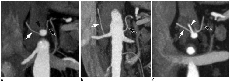
Fig. 5. CT angiography MIP images show right inferior phrenic artery (RIPA, white arrow) and left inferior phrenic artery (LIPA, black arrow) originating separately without truncus.
A. Both RIPA and LIPA originate from celiac axis (black arrowhead). B. RIPA originates from right renal artery and LIPA from aorta. C. RIPA originates from left gastric artery (white arrowhead) and LIPA from celiac axis (not shown). MIP = maximum intensity projections

