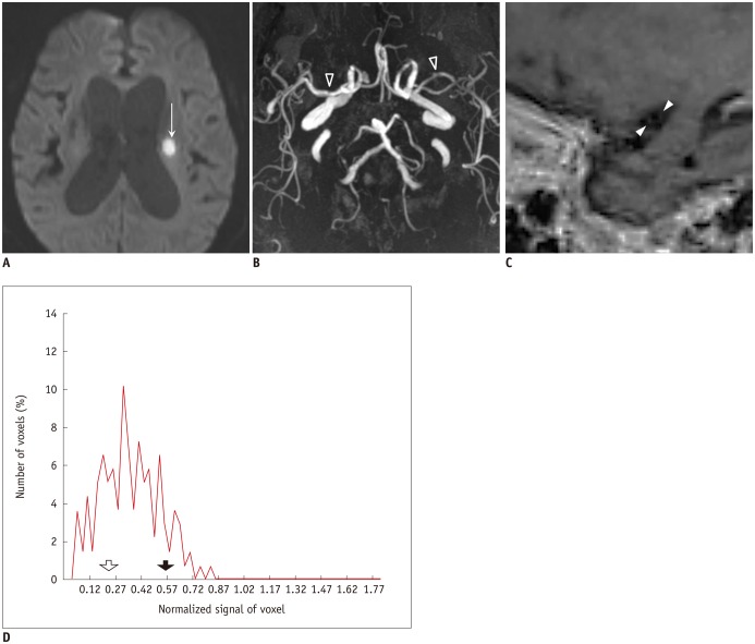
Fig. 4. Case of SAD.
86-year-old woman visited emergency room for dysarthria. DWI showed small acute ischemic stroke lesion in left periventricular white matter (arrow on A). Minimal irregular contour (arrowheads) of M1 segment of left MCA was visible (B), and degree of stenosis was 20%. On sagittal image (C), enhancement score was 0. Histogram analysis of lesion side M1 (D) showed low 90P (solid arrow, 0.568) and low GM (empty arrow, 0.256) in lesion side M1. DWI = diffusion-weighted image, GM = geometric means, MCA = middle cerebral artery, SAD = small artery disease, 90P = 90th percentile

