
Figure 1.
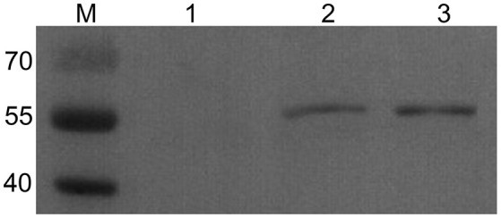
Western blot analysis of TgSPATR expressed in HEK293. M: marker 40–70 kDa; (1) negative control, the lysate of HEK293 transformed with empty pVAX1 vector; (2) the lysate of HEK293 transformed with pVAX1-TgSPATR; (3) purified rTgSPATR of T. gondii expressed in E. coli.