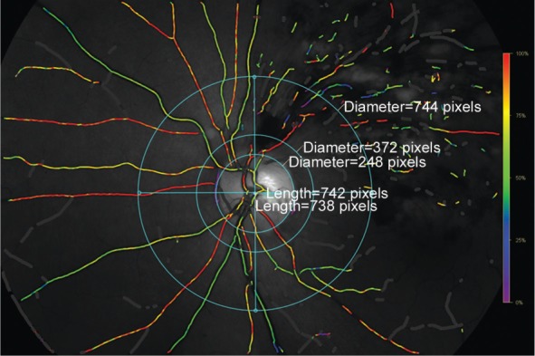
Figure 1. Fundus photograph showing the retinal vessels as imaged and measured by the oximeter.

In the superior, inferior, nasal and temporal quadrant, we selected the thickest arteriole and thickest venule for each quadrant between the two circles outside of the optic disc border.
