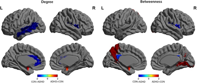
Figure 3.
Differences between attention-deficit hyperactivity disorder (ADHD) and Control (CON) groups in regional degree and betweenness. Regions with significant group differences in regional degree (left) and betweenness (right) for networks thresholded at minimum density of full connectivity, overlaid on ICBM152 surface template. The color bar represents log(1/P-value). Hot colors indicate regions with higher degree or betweenness in ADHD compared with Controls, while cold colors indicate regions with higher degree or betweenness in Controls compared with ADHD. ADHD had greater nodal degree in the bilateral amygdalae, and reduced degree in left (L) anterior cingulate, L mid temporal pole and right (R) rolandic operculum. Nodal betweenness was increased in ADHD compared with Controls in L amygdala and precuneus, and R lingual gyrus, but reduced in L posterior cingulum and R rolandic operculum.

