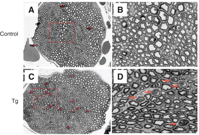
FIGURE 7.

Axon degeneration in the peripheral nervous system of NCmahTg mice. Light microscope images of cross-sections of toluidine blue-labeled sciatic nerves from control (wild-type or Cre-positive (no Cmah transgene)) mice (A and B) and NCmahTg mice (C and D) revealed increased degeneration (arrows) in the NCmahTg mice. Boxed areas in A and C are shown at higher magnification in the adjacent panels (B and D). Data are representative of the results obtained from two (control) or three (NCmahTg) mice.
