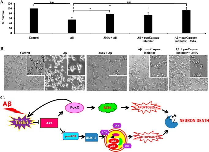
FIGURE 8.
Contribution of both autophagy and apoptosis in Aβ-induced neuronal cell death. A, graphical representation of cell survival following Aβ treatment with pan-caspase inhibitor and 3-methyladenine (3-MA), or both, for 16 h. Data are represented as mean ± S.E. of three independent experiments. *, p < 0.05; **, p < 0.001. B, representative phase contrast micrographs of neuronal PC12 cells before and after treatment of Aβ for 16 h. C, Trib3 is up-regulated upon Aβ exposure, Trib3 then interacts with Akt and inhibits its phosphorylation, as a result of which two molecules are affected. On the one hand, FoxO gets activated, translocates to the nucleus, and causes transcription of the pro-apoptotic protein Bim; FoxO also binds to the promoter region of Trib3 amplifying the feed-forward loop between them. On the other hand, mTOR gets inhibited, as a result of which Ulk1 is activated which then initiates the autophagic cascade as seen by the induction of cleavage of LC3 and increased accumulation of p62. Trib3 thus designs death of neurons by both autophagy and apoptosis.

