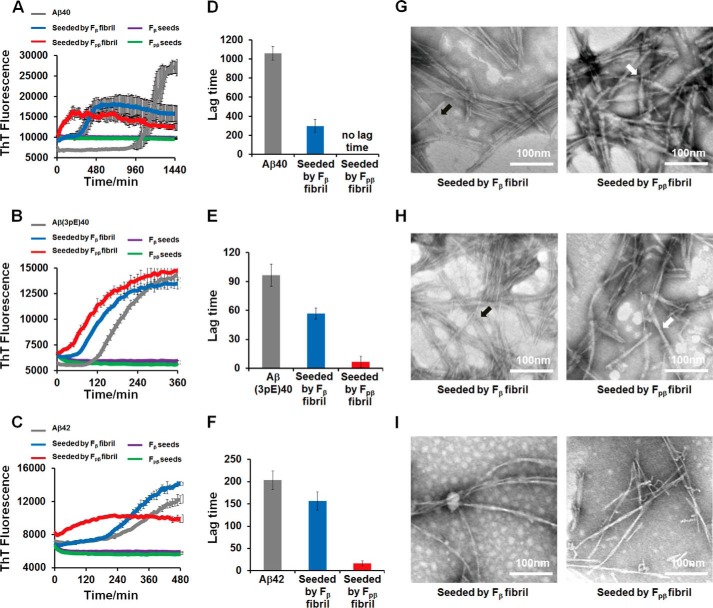
FIGURE 6.
Propagation of Fpβand Fβ fibrils in vitro was monitored by ThT kinetics assay and TEM images. Propagation of preformed 10% Fpβ (red curves) and Fβ fibrils (blue curves) in 40 μm Aβ40 (A), 10 μm Aβ(3pE)40 (B), and 10 μm Aβ42 (C) in vitro at pH 7.4 was monitored by ThT fluorescence kinetics assay. The lag phase for Aβ40 (D), Aβ(3pE)40 (E), and Aβ42 (F) showed that both Fpβ (red column) and Fβ (blue column) can shorten the lag phase for amyloid formation. The structural natures of Aβ40, Aβ(3pE)40, and Aβ42 aggregates after seeding were examined using TEM (G, H, and I, respectively); left and right panels are structural images of Fβ-seeded fibrils and Fpβ-seeded fibrils, respectively. The TEM samples were prepared after 24-h incubation. For Aβ40 (G) and Aβ(3pE)40 (H), the structural information of parental Fβ fibrils and Fpβ fibrils can be inherited. The black and white arrowheads point to ribbon-like and twisted cylindrical morphologies, respectively. However, Aβ42 cannot sample the structural information from either Fβ or Fpβ fibrils (I). ThT fluorescence kinetics of different soluble Aβ species without any seeds and Fβ or Fpβ seeds only were also conducted as controls. Error bars represent S.E. (n = 3 independent measurements).

