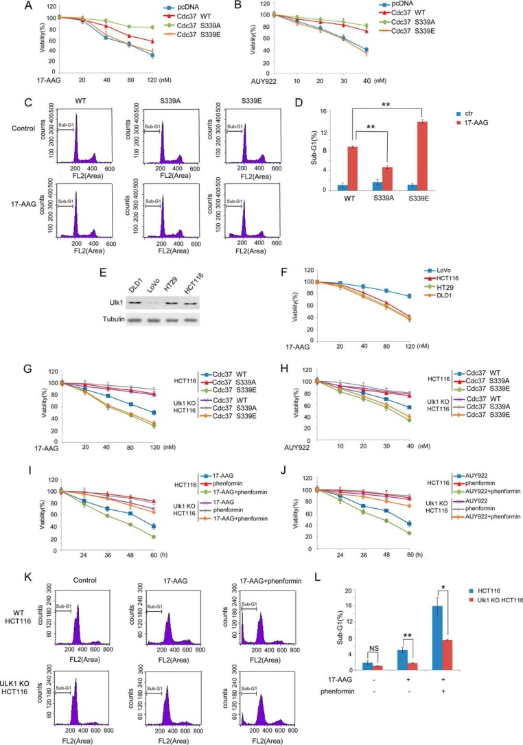
FIGURE 6.
Ulk1 sensitized cancer cells to Hsp90 inhibitor. A and B, a set of RNAi-resistant rescue forms of Cdc37 plasmids was transfected into stable Cdc37-RNAi HCT116 cells. Cells were then treated with 17-AAG (A) or AUY922 (B) at different concentration for 36 h, and cell proliferation was tested with the MTT assay. C and D, a set of RNAi-resistant rescue forms of Cdc37 plasmids were transfected into stable Cdc37-RNAi HCT116 cells. Cells were treated with 100 nm 17-AAG for 72 h, and the cells were then harvested for flow cytometry analysis. The percentage of sub-G1 was shown in D. E, Western blotting was performed to determine Ulk1 expression inhuman colon cancer cell lines DLD1, LoVo, HT29, and HCT116. F, DLD1, LoVo, HT29, and HCT116 cells were treated with 17-AAG at different concentration for 36 h, and cell proliferation was tested with the MTT assay. G and H, a set of the RNAi-resistant rescue form of Cdc37 plasmids was transfected into Cdc37-KD or Cdc37-KD/Ulk1-KO HCT116 cells. Cells were then treated with 17-AAG (G) or AUY922 (H) at different concentration for 36 h, and cell proliferation was tested with the MTT assay. I and J, Wild-type and Ulk1-KO HCT116 cells were treated with 40 nm 17-AAG (I) or 20 nm AUY922 (J) combined with phenformin, and cell proliferation was tested with the MTT assay. K and L, wild-type and Ulk1-KO HCT116 cells were treated with 100 nm 17-AAG combined with phenformin for 72 h, and the cells were then harvested for flow cytometry analysis. The percentage of sub-G1 was shown in L. NS, not significant. *, p < 0.05; **, p < 0.01.

