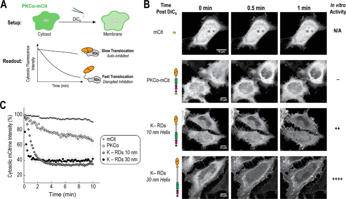
FIGURE 7.
Disrupting PKCα autoinhibition increases membrane translocation. A, upon stimulation with 0.05 mg/ml of DiC8 lipid, HeLa cells were imaged for 10 min at 22 °C using a confocal microscope. Movies were analyzed by selecting and monitoring fluorescence cytosolic intensity over time for three cytosolic regions per cell. B, representative images and C, analysis of one field of cells per condition (mean ± S.E., n ≥ 2 cells). For each condition, at least two independent experiments were performed and in total more than four cells per condition were analyzed. Autoinhibition was increasing disrupted by introducing either 10- or 30-nm ER/K linkers between the catalytic domain and regulatory domains, as previously described (20). The rate of translocation was compared with mCitrine-labeled full-length PKCα and a mCitrine (mCit) fluorophore control.

