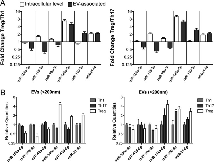
FIGURE 4.
CD4+ T cell subset-specific EV-associated miRNA signature does not mirror differences among subsets at the intracellular level. A, histogram graphs (means with S.E.) showing the fold change (evaluated by RT-qPCR and normalized relative to global mean) of the indicated extracellular miRNAs when comparing Treg with either Th1 (left panel) or Th17 (right panel) at the intracellular level (in ex vivo purified resting cells, white) compared with EV-associated level (gray). B, column bars (mean with S.E.) showing the relative quantities (evaluated by RT-qPCR and normalized relative to global mean) of the indicated extracellular miRNAs in smaller (<200 nanometers, left panel) versus larger (>200 nanometers, right panel) vesicles. The results were obtained by nine biological replicates.

