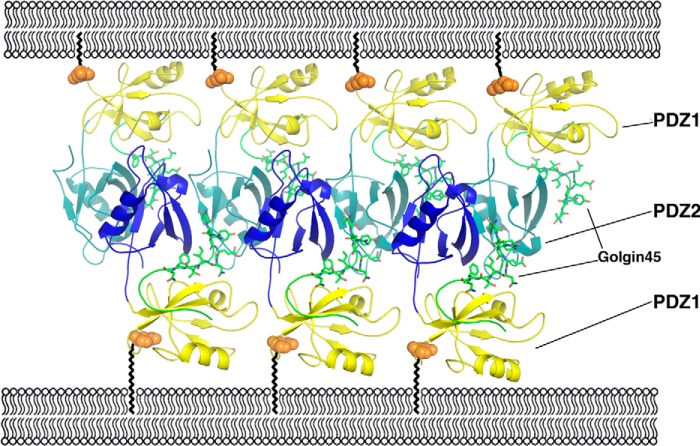
FIGURE 6.
Molecular models of GRASP55-Golgin45-mediated mid-cisternae membrane stacking. PDZ1 domains are colored yellow, whereas PDZ2 domains of GRASP55 are colored blue. Golgin45 C-terminal peptides are shown with a stick model and colored green. The ball and zigzag lines represent GRASP55 N-terminal myristoylation. A series of GRASP55 and Golgin45 molecules form ordered membrane-associated protein arrays between two apposing membranes.

