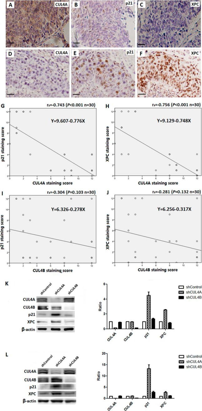
FIGURE 5.
Inverse correlation of CUL4A and P21 or XPC expression in SCC patients. A–F, representative staining of CUL4A, P21, and XPC in SCC specimens. Regression line plots showed significant inverse correlation of P21 or XPC with CUL4A (G and H) but not CUL4B (I and J). K and L, depletion of CUL4A resulted in increased accumulation of XPC and P21 in NCI-H520 SCC cells (K) or NCI-H446 SCLC cells (L), as determined by immunoblotting using antibodies again CUL4A, CUL4B, and β-actin (internal loading control). The levels of CUL4A, CUL4B, XPC, and P21 (relative to that of β-actin) in the shCUL4A and shCUL4B cell lines were quantitatively determined using Odyssey and plotted against those of the shControl (right panels).

