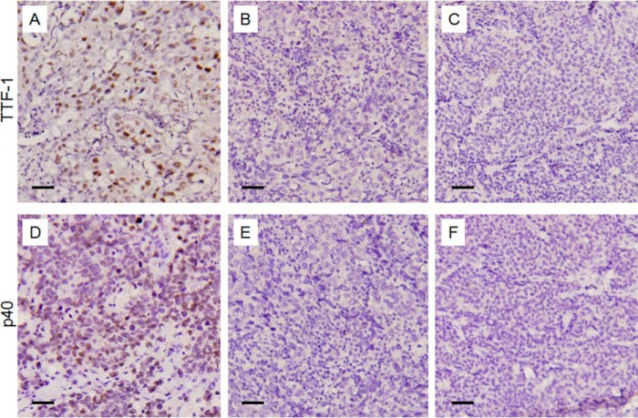
FIGURE 6.
Negative IHC staining of nuclear TTF-1 and P40 for diagnosis of large cell lung carcinomas. A, representative staining of TTF-1 in adenocarcinoma as positive control. B and C, IHC staining of TTF-1 in two representative large cell lung carcinoma cases. D, representative staining of P40 in squamous cell lung carcinoma as positive control. E and F, IHC staining of P40 in the corresponding specimens as B and C.

