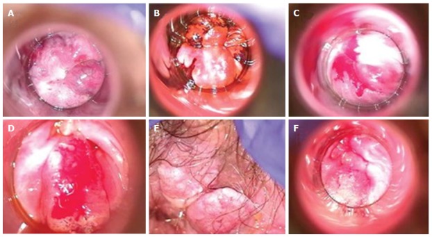
Figure 2.

High-resolution anoscopy of representative examples of anal intraepithelial lesions. A: Low grade AIN lesion after acetic acid application with representative acetowhitening; B: Low grade AIN lesion after application of Lugol’s iodine with brown area representing normal uptake by glycogenated cells, and “mustard” colored area representing negative uptake and suggestive of dysplasia; C: High grade AIN seen after application of acetic acid and the dense acetowhite change; D: High grade AIN with concern for invasion; E: External/perianal high grade AIN after application of acetic acid; F: High grade AIN with concern for invasion. Reproduced with permission[85]. AIN: Anal intraepithelial neoplasia.
