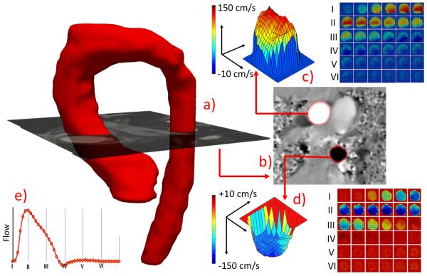
Figure 1.
(a) MRA reconstructed aorta with superimposed PC-MRI magnitude image plane depicting the flow hemodynamic computation locations. (b) Corresponding phase image with segmented ascending and descending aortic lumens for flow hemodynamic quantification (c and d) and reconstructed flow-wave form (e).

